Difference Between Scanning and Transmission Electron Microscope
Transmission electron microscope TEM uses electrons which pass through the sample so the resulting micrograph image shows everything within the sample in black and white for example organelles in a cell. The main difference between scanning and transmission electron microscopy is that scanning electron microscopy generates A.

Transmission Electron Microscope Tem Micrograph Showing Several Peripheral Myelinated Fibers And A Electron Microscope Microscopy Electron Microscope Images
Choose From 1000s Of Microscopes.

. Scanning electron microscope SEM Compound microscope. During the scanning the beam loses energy in different amounts according to the surface it is on. 3 00 000 times.
An electron beam is directed onto the sample to be magnified and some. In this review we consider specifically contributions and limitations of STEM for the investigation of amyloid assembly pathways fibril polymorphisms and structural models of amyloid fibrils. Transmission Electron Microscope TEM Fig.
4 08 000 times. Transmission electron microscope TEM Answer. 10 rows The transmission electron microscopy TEM principle as the name suggests is to use the.
In my previous notes I have already discussed Transmission Electron Microscope and Scanning Electron Microscope their working principle parts definition application advantages disadvantages and light path. 6 rows A scanning electron microscope only scans a specimen. 20 Difference Between Transmission and Scanning Electron Microscope.
You can check them out. A scanning electron microscope doesnt use a concentrated electron beam to penetrate the object as a transmission electron microscope does. The magnification power of an optical microscope is.
The main difference between Scanning Electron Microscope and Transmission Electron Microscope is that scanning electron microscopes produce surface images by reflecting electrons from the specimens surface while transmission electron microscopes produce an internal image of the specimen by emitting electrons that cross through it. Scanning transmission electron microscopy STEM is often used to delineate the assembly mechanism and structural properties of amyloid aggregates. 4 12 000 number of times.
Difference between Transmission Electron Microscope TEM and Scanning Electron Microscope SEM The main difference between a transmission electron microscope TEM and scanning electron microscope is the fact that TEM operates by penetrating electrons through the specimen and producing image by capturing the emitted electrons from the. TEM Transmission Electron Microscope and. 2 09 000 no.
Differences between Transmission Electron Microscope and Scanning Electron Microscope-By focusing and magnifying the mainly internal ultrastructure of the cell the Transmission Electron Microscope utilizes electromagnets as a lensHowever the Scanning Electron Microscope was utilized for a detailed investigation of the surface of a particular. Under an electron microscope you can magnify an object up to. A scanning electron microscope doesnt use a concentrated electron beam to penetrate the object as a transmission electron microscope does.
High-Quality Microscopes At The Lowest Cost. Difference between scanning electron microscopy sem and. Ad Get The Lowest Price On Our Wide Selection Of Compound Microscopes.
A high-resolution image that reveals detailed intracellular objects. The two most common types of electron microscopes are transmission TEM and scanning SEM systems but the differences between these two instruments can be fairly nuanced. Transmission electron microscope tem introduction to.
During the scanning the beam loses energy in different amounts according to the surface it is on. Here we hope to provide a fundamental primer for individuals looking to get started with this powerful technique. 25 Years of Industry Experience.
Interaction electron beam with sampleThe Scanning Electron Microscope SEM produces images by probing the specimen with a focused electron beam that is scanned across a rectangular area of the specimen raster scanning. Scanning transmission electron microscopy springerlink. This limits the amount of information.
There are two families of electron gunsConventional thermionic emitters. The Only Company Offering TEM in Compact Benchtop Formats. Scanning Electron Microscope SEM Imagine you are in a dark room.
A high-resolution image that appears three-dimensional. Request a Quote Now. A transmission electron microscopes TEM can magnify a sample up to one million times.
Ad Shop Electron Microscopes for Life Material Sciences. 21 rows There are two types of electron microscopes. Instead it scans a beam across the object.
The sample must be cut extremely thin. Instead it scans a beam across the object. A low-resolution image that appears three-dimensional.
Optical microscope scanning electron microscope sem. An introduction to electron microscopy stem bines.

Differences Between Light Microscope And Electron Microscope Electron Microscope Electrons Microscopic

The Cell An Image Library Image Cil 11397 Microscopic Photography Electron Microscope Images Photosynthesis And Cellular Respiration

Chapter 3 Section 1 Microscopes Cells Under The Microscope Objectives Describe How Scie Scanning Electron Microscope Optical Microscope Microscope Objective
Schematic Diagram Of A Transmission Electron Microscope B Scanning Electron Microscope Scanning Electron Microscope Electron Microscope Microscopic Images

How Scanning Electron Microscopes Work Scanning Electron Microscope Electron Microscope Scanning Electron Microscopy

Golgi Body Yale Histology Gallery Microscopic Photography Electron Microscope Microscope Art

Picture Fun Science Electron Microscope Science

There Are Two Categories Of Microscopes Based On The Principle On Which Magnificati Scanning Electron Microscope Electron Microscope Electron Microscope Images
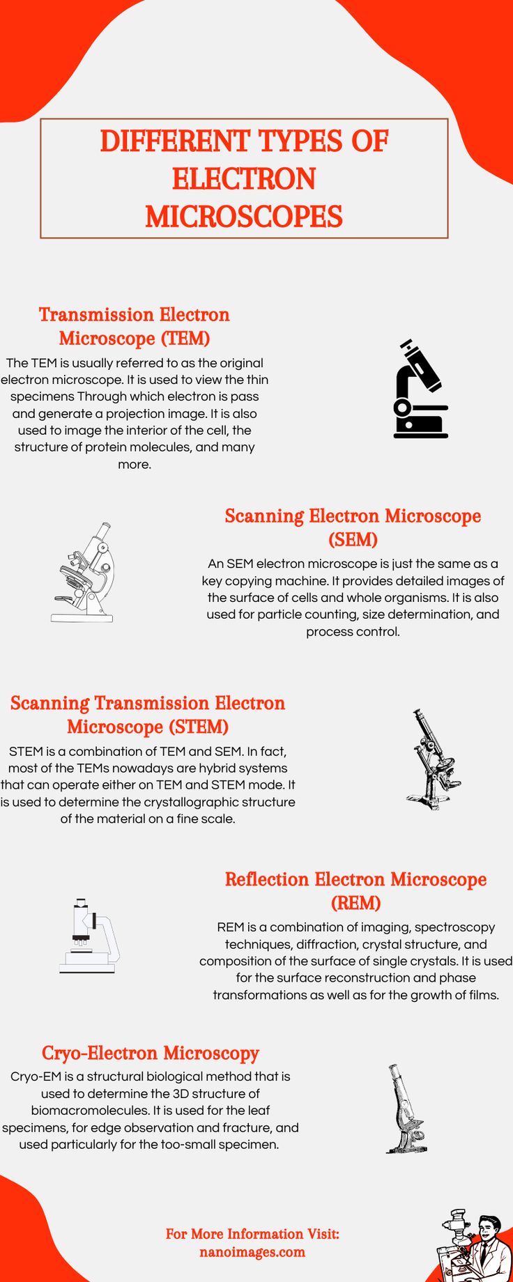
Different Type Of Electron Microscopes Electron Microscope Scanning Electron Microscope Electrons
![]()
Labelled Artwork Showing How A Transmission Electron Microscope Tem Works Electron Microscope Scanning Electron Microscope Cell Organelles

Figure 12 These Schematic Illustrations Compare The Components Of Transmission Electron Microsco Microbiology Scanning Electron Microscope Electron Microscope

Bacteria And Fungus Seen In The Scanning And Transmission Electron Microscope Scientific Photography Microorganisms Food Animals Electron Microscope
![]()
Transmission Electron Microscopy Microscopy Electrons Scanning Electron Microscope

Hf 3300 300 Kv Fe Tem Hitachi High Technologies America Inc Electron Microscope Medical Lab Technician Transportation Design

The Mitochondria Is Like The Digestive System Of The Cell It Process Nutrients And Creates E Microscopic Photography Scanning Electron Micrograph Mitochondria

Wellcome Education On Twitter Microscopic Images Electron Microscope Images Scanning Electron Micrograph

Golgi Apparatus Tem Cell Organelles Macro And Micro Animal Cell

Which Microscope Learning Science Biology Resources Teaching Biology

Visualization Of The Cell Using Em Scanning Electron Microscope Microscopic Photography Microscopy


Comments
Post a Comment